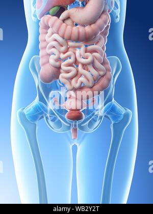
Abdominal Anatomy Pictures Female - Normal Abdominal Anatomy Medical Illustration - The abdominal cavity is the part of the body that houses the stomach, liver, pancreas, kidneys, gallbladder, spleen, and the large and small intestines.the diaphragm marks the top of the abdomen and the horizontal line at the level of the top of the pelvis marks the bottom.
Abdominal Anatomy Pictures Female - Normal Abdominal Anatomy Medical Illustration - The abdominal cavity is the part of the body that houses the stomach, liver, pancreas, kidneys, gallbladder, spleen, and the large and small intestines.the diaphragm marks the top of the abdomen and the horizontal line at the level of the top of the pelvis marks the bottom.. The diaphragm forms the upper surface of the abdomen. At the level of the pelvic bones, the abdomen. Huge collection, amazing choice, 100+ million high quality, affordable rf and rm images. Sterilization, removal of female ovarian organs. See abdominal organs stock video clips.
We're going to take apart a plastic anatomy model and see what we can find in the abdomen. In this anatomy course you will explore the organs involved in our food digestion and discover the common causes of abdominal and pelvic pain. Browse 2,011 female urinary system stock photos and images available, or search for lungs to find more great stock photos and pictures. Abdominal anatomy pictures female : The front of the body is at right.
Anatomy Of The Female Abdomen And Pelvis Stock Photo Alamy from l450v.alamy.com There are two hip bones, one on the left side of the body and the other on the right. Select from premium abdominal anatomy of the browse 7,978 abdominal anatomy stock photos and images available, or search for abdominal muscles or human body part to find more great stock. Together, they form the part of the pelvis called the pelvic girdle. Female abdominal anatomy pictures, download this wallpaper for free in hd resolution. It is present near the semilunar line of the transversus abdominis muscle. Spleen is 1 inch thick, 3 inches broad and 5 inches long. Browse 2,177 female pelvis stock photos and images available, or search for female pelvis illustration to find more great stock photos and pictures. Learn vocabulary, terms and more with flashcards, games and other study tools.
Find the perfect abdominal hernia stock photo.
Über 7 millionen englischsprachige bücher. Female abdominal anatomy pictures, download this wallpaper for free in hd resolution. We'll identify as many organs as we can, see how they fit into the. The major organs of the abdomen include the small intestine, large intestine, and stomach. Spigellian hernia can be congenital. The abdomen (commonly called the belly) is the body space between the thorax (chest) and pelvis. Find the perfect abdominal hernia stock photo. The liver, stomach, and abdominal contents are clearly identified and labeled, including the cecum, ascending colon, transverse colon, descending colon, and small intestine. These are often rare and complex types of hernia that may be difficult to diagnose due to their location and non. Together, these three turn nutrients into usable energy, as well as help dispose of solid waste. Connective tissue called the mesentery holds the abdominal organs together. Veterinary services for the treatment of pets. Select from premium abdominal anatomy of the browse 7,978 abdominal anatomy stock photos and images available, or search for abdominal muscles or human body part to find more great stock.
Spigellian hernia can be congenital. Spleen is the biggest lymphoid organ present in the upper far left portion of the abdomen in the left hypochondrium and is surrounded by peritoneum. He has been with healthiack.com since 2012 and has written and reviewed well over 500 coherent articles. See abdominal muscle stock video clips. Together, they form the part of the pelvis called the pelvic girdle.
Female Abdominal Anatomy Artwork Photograph By Peter Gardiner from images.fineartamerica.com Browse 2,177 female pelvis stock photos and images available, or search for female pelvis illustration to find more great stock photos and pictures. Learn vocabulary, terms and more with flashcards, games and other study tools. No need to register, buy now! Spleen is 1 inch thick, 3 inches broad and 5 inches long. Anatomy of stomach artery 12 photos of the anatomy of stomach artery anatomy gastric artery, anatomy of left gastric artery, anatomy of right gastric artery, human anatomy, anatomy gastric artery, anatomy of left gastric artery, anatomy of right gastric artery Dreamstime is the world`s largest stock photography community. Spleen is the biggest lymphoid organ present in the upper far left portion of the abdomen in the left hypochondrium and is surrounded by peritoneum. We'll identify as many organs as we can, see how they fit into the.
Browse 2,011 female urinary system stock photos and images available, or search for lungs to find more great stock photos and pictures.
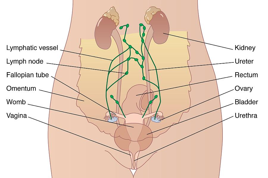
Sterilization, removal of female ovarian organs. The abdomen contains all the digestive organs, including the stomach, small and large intestines, pancreas, liver, and. The abdomen (commonly called the belly) is the body space between the thorax (chest) and pelvis. We'll identify as many organs as we can, see how they fit into the. Anatomy of the trunk with heart, kidneys and bladder. Don't forget to share this picture with others via facebook, twitter, pinterest or other social medias! Veterinary services for the treatment of pets. Vertebral disorder human body with internal organ human bowel pain body organs heart lung intestine organ of the human body constipation intestine human body and internal organs the digestive tract gut. Together, they form the part of the pelvis called the pelvic girdle. The enlargement of spleen is referred to as splenomegaly. Female abdominal anatomy pictures, download this wallpaper for free in hd resolution. Together, these three turn nutrients into usable energy, as well as help dispose of solid waste. We also distinguish the vena cava inferior and the abdominal aorta.
We're going to take apart a plastic anatomy model and see what we can find in the abdomen. The enlargement of spleen is referred to as splenomegaly. The abdomen (commonly called the belly) is the body space between the thorax (chest) and pelvis. It is a type of rare abdominal wall defect characterized by a protrusion in the abdominal wall that comprises preperitoneal fat, omentum, or an organ. Huge collection, amazing choice, 100+ million high quality, affordable rf and rm images.
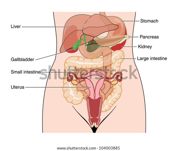
Drawing Abdomen Showing Abdominal Organs Female Stock Illustration 104003885 from image.shutterstock.com He has been with healthiack.com since 2012 and has written and reviewed well over 500 coherent articles. The abdomen (commonly called the belly) is the body space between the thorax (chest) and pelvis. The image also shows the pelvis, uterus, and urinary. Sterilization, removal of female ovarian organs. Female abdominal anatomy pictures, download this wallpaper for free in hd resolution. In this anatomy course you will explore the organs involved in our food digestion and discover the common causes of abdominal and pelvic pain. Vertebral disorder human body with internal organ human bowel pain body organs heart lung intestine organ of the human body constipation intestine human body and internal organs the digestive tract gut. Dreamstime is the world`s largest stock photography community.
No need to register, buy now!
The image also shows the pelvis, uterus, and urinary. These are often rare and complex types of hernia that may be difficult to diagnose due to their location and non. Female abdominal anatomy pictures was posted in may 15, 2015 at 9:16 am. Don't forget to share this picture with others via facebook, twitter, pinterest or other social medias! The abdominal cavity is the part of the body that houses the stomach, liver, pancreas, kidneys, gallbladder, spleen, and the large and small intestines.the diaphragm marks the top of the abdomen and the horizontal line at the level of the top of the pelvis marks the bottom. Together, they form the part of the pelvis called the pelvic girdle. We're going to take apart a plastic anatomy model and see what we can find in the abdomen. Spleen is 1 inch thick, 3 inches broad and 5 inches long. Together, these three turn nutrients into usable energy, as well as help dispose of solid waste. Veterinary services for the treatment of pets. Find the perfect abdominal hernia stock photo. It is a type of rare abdominal wall defect characterized by a protrusion in the abdominal wall that comprises preperitoneal fat, omentum, or an organ. Sterilization, removal of female ovarian organs.


0 Komentar